When your pet needs surgery, it’s natural to feel a mix of emotions—concern, confusion, and the deep desire to ensure their safety and comfort. One of the most critical, yet often overlooked, aspects of surgical success is diagnostic imaging, specifically pet ultrasound and pet X-ray. These tools serve as a window into your pet’s body, offering vital information that helps veterinarians make accurate diagnoses, plan procedures, and prevent complications.
Thanks to advances in veterinary medicine, both the safety and efficacy of pet surgery have significantly improved, with modern protocols and technology ensuring better outcomes for pets.
In this blog, we’ll take a closer look at how pet ultrasound and X-ray imaging are used before both elective and urgent pet surgery procedures, what pet owners in Doha should know, and how these technologies contribute to better outcomes for your furry companion.
Why Diagnostic Imaging Is Essential Before Surgery
Ultrasound and pet X-ray are not just technologies—they are essential diagnostic tools. Whether your pet is scheduled for a routine procedure or a life-saving operation, these imaging methods help answer crucial questions such as:
What exactly is causing your pet’s symptoms?
Where is the affected organ, mass, or blockage located?
Is your pet healthy enough to undergo surgery?
How can the surgery be performed with the least amount of risk?
Can imaging identify potential complications before surgery?
Imaging helps veterinarians assess your pet's health and current condition, guiding surgical decisions and ensuring the best possible outcome for your pet's well-being.
Without this information, even experienced veterinarians would be operating with limited visibility—something no pet owner would want.
Understanding X-Rays in Pre-Surgical Planning
What Is an X-Ray?
An X-ray, or radiograph, uses low levels of electromagnetic radiation to create images of internal structures. It is especially effective at showing bones, joints, the chest cavity, abdomen, and dense foreign objects.
When Are X-Rays Used Before Surgery?
The Role of Pet Ultrasound & Pet X-Ray Before Pet Surgeries 1
X-rays are often the first step in the diagnostic process and are particularly useful for:
Identifying broken bones or joint dislocations
Detecting tumours or growths
Locating swallowed objects like stones, toys, or bones
Detecting a foreign body in pets, such as ingested items that may cause blockages
Examining the lungs and heart before anaesthesia
Evaluating spinal problems or arthritis
Finding bladder or kidney stones
Hip X-rays may require sedation or special positioning to ensure accurate results, as these procedures can be more challenging and require the animal to remain calm.
Let’s say your dog swallowed a sock or your cat isn’t eating and seems to be in pain. An X-ray can reveal if there’s a blockage or foreign object that may require surgical removal.
Benefits of X-Rays
Quick and non-invasive
Helps avoid exploratory surgery
Gives a clear view of bones and dense tissues
Useful in emergencies
However, X-rays have limitations when it comes to soft tissues, which is where pet ultrasound comes into play.
The Importance of Ultrasound in Soft Tissue Evaluation
The Role of Pet Ultrasound & Pet X-Ray Before Pet Surgeries 2
What Is an Ultrasound?
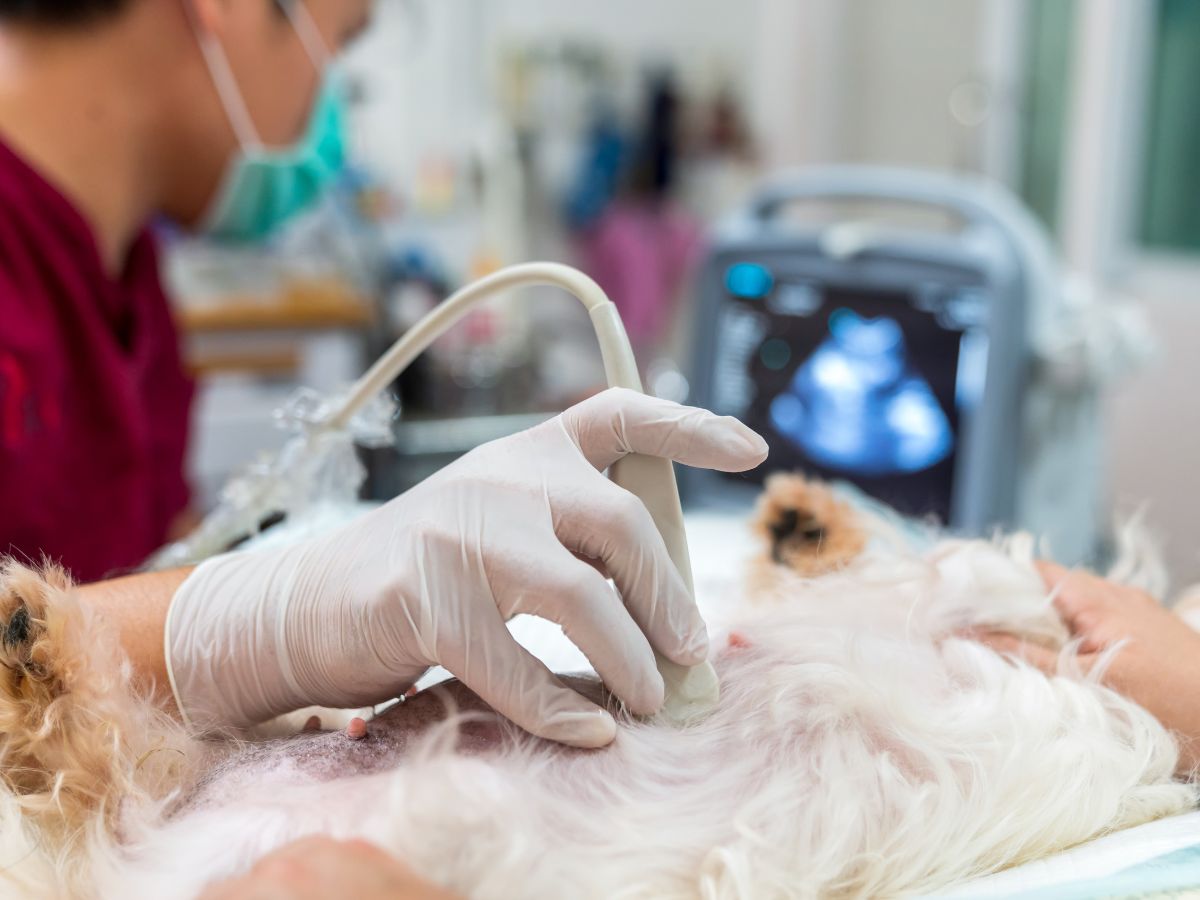
Ultrasound imaging uses high-frequency sound waves to create real-time images of your pet’s internal organs. It is ideal for examining soft tissues, such as the liver, kidneys, bladder, uterus, intestines, heart, and the gastrointestinal tract.
An abdominal ultrasound is commonly used to detect abnormalities or obstructions in the abdomen, including issues within the gastrointestinal tract. Ultrasound can also assess blood flow in organs and tissues using Doppler technology, providing valuable information about circulation.
Unlike X-rays, ultrasounds can capture movement, allowing vets to see how organs function, if there’s fluid buildup, or if a mass is solid or cystic.
When Is Ultrasound Used Before Surgery?
Ultrasound is often used to:
Assess the heart before anaesthesia in pets with cardiac issues, and diagnose heart disease using echocardiograms or cardiac ultrasound
Determine the origin of abdominal pain
Evaluate tumours or abnormal masses, including assessment of lymph nodes for abnormalities or cancer
Identify organ enlargement, damage, or inflammation
Locate fluid accumulation in the chest or abdomen
Aid in biopsies or fluid sampling (ultrasound-guided)
Perform pregnancy diagnosis in pets
For example, if your cat has a swollen abdomen or your dog has recurring vomiting, an ultrasound can help pinpoint whether there’s a gastrointestinal blockage, liver problem, or an issue with the kidneys.
Benefits of Ultrasound
Painless and non-invasive
No radiation exposure
Allows dynamic, real-time observation
Can guide needle biopsies with precision, helping to target abnormal tissue for sampling
Ultrasound is especially valuable for internal organ issues that cannot be easily detected through physical exams or X-rays.
Combining Ultrasound and X-Ray for Accurate Diagnosis
While both ultrasound and X-ray are powerful individually, they are often used together for a comprehensive diagnosis. Advanced imaging techniques like digital ultrasound and radiography contribute to a more accurate pet's diagnosis by providing clearer and more detailed images. Here’s why:
X-rays offer a good overview of the chest and skeletal structure
Ultrasound provides detailed insight into soft tissue structures and fluid
When combined, they help your vet confidently plan the safest and most effective surgical approach
For example, in a pet with suspected cancer, an X-ray may reveal a suspicious mass near the ribs, and an ultrasound can help determine its nature, size, and whether it has spread to nearby organs.
How Imaging Reduces Surgical Risks
1. Pre-Anaesthetic Evaluation
Before surgery, especially in older pets or those with known health conditions, your vet will want to assess the lungs, heart, and abdominal organs to determine if the pet is a good candidate for anaesthesia. Imaging plays a key role in this evaluation.
2. Planning the Incision and Procedure
Surgery should never be guesswork. Imaging allows the vet to know exactly where to operate, how large the incision should be, and what complications to anticipate. This reduces time under anaesthesia and improves recovery outcomes.
3. Avoiding Unnecessary Surgery
Sometimes, diagnostic imaging reveals that surgery may not be needed at all. Conditions such as inflammation, infections, or benign cysts might be treated with medication or minimally invasive methods instead.
What Pet Owners in Doha Should Expect
As a pet owner, it’s important to understand the diagnostic process and be prepared to authorise imaging procedures before surgery. In Doha, veterinary clinics like Royal Vet Clinic offer advanced imaging services as part of their pre-operative protocol.
Here’s what to expect:
A consultation and physical exam
A recommendation for X-ray, ultrasound, or both
An explanation of what the imaging will show
Sedation if needed (especially for ultrasound or if the pet is anxious)
Immediate or same-day results
A surgical plan based on findings
You’ll also have the opportunity to ask questions about your pet’s condition and how the surgery will proceed.
Frequently Asked Questions
Q. Is diagnostic imaging safe for my pet?
Yes. X-rays use minimal radiation and are safe when used appropriately. Ultrasounds involve no radiation at all. In some cases, mild sedation may be used to keep pets still during imaging.
Q. How long does the imaging process take?
X-rays typically take 10–15 minutes. Ultrasounds may take 20–30 minutes, depending on the complexity and whether a detailed scan or biopsy is required.
Q. Will my pet need to fast before an ultrasound or X-ray?
Usually yes, especially if sedation is required. Both cats and dogs may need special fasting or preparation instructions, so your vet will advise you on the best approach before the appointment.
Q. What can imaging detect in my pet?
Imaging helps identify a wide range of health issues, including soft tissue problems, gastrointestinal, heart, and nervous system conditions. It is also valuable for diagnosing urethral blockages that may require surgical removal, as well as other internal abnormalities.
Q. What happens if the imaging shows something serious?
If a mass, blockage, or organ problem is detected, your vet will discuss surgical or medical options and provide a treatment plan tailored to your pet’s condition. For urethral blockages, your vet may recommend surgery to remove the obstruction and will discuss post-operative care to ensure normal urination.
Q. Can imaging be done the same day as the surgery?
In emergencies, yes. However, planned surgeries usually involve imaging at least a day before to allow time for planning and lab tests.
Q. Is imaging covered in the cost of surgery?
This depends on the clinic. At Royal Vet Clinic, we offer transparent pricing and package options that include pre-operative imaging, lab work, and post-operative care.
Q. How is recovery after surgery for most pets?
Most pets, including both cats and dogs, recover well after surgery with proper care and monitoring at home. Your vet will provide specific post-operative instructions to help ensure a smooth recovery.
The Role of the Pet Owner in Surgical Success
Your cooperation plays a big role in successful surgical outcomes. Ensure you follow your vet’s instructions for:
Fasting before imaging or surgery
Providing a complete medical history
Discussing any ongoing medications
Monitoring your pet post-imaging and post-surgery
Don’t hesitate to ask questions about the results or the reasons behind the imaging tests. A well-informed pet parent is an empowered one.
Why Choose Royal Vet Clinic in Doha for Imaging and Surgery?
At Royal Vet Clinic, we are committed to the highest standards of veterinary medicine. We provide comprehensive veterinary care, including advanced diagnostics and surgery. Our facility is equipped with state-of-the-art pet ultrasound and X-ray technology, allowing us to make accurate diagnoses and perform precise, minimally invasive surgeries.
While ultrasound is excellent for imaging soft tissues, it cannot pass through bone to visualise the brain and spinal cord. For detailed imaging of the brain and spinal cord, advanced diagnostics such as CT or MRI scans are required.
Our experienced veterinary team works closely with pet owners to ensure they understand each step of the process. From initial consultation to post-surgery recovery, your pet receives compassionate, professional care tailored to their individual needs.
In addition to surgical diagnostics, we also provide complete pet dental care services for pets, ensuring every part of your furry companion’s health is well looked after.
If your pet is facing surgery or showing signs of illness, don’t wait. Visit Royal Vet to book a consultation or learn more about our imaging and surgical services. We’re here to help you make informed decisions that lead to healthier, happier pets.
Traveling with pets is a growing trend among families in Doha, especially those relocating to Europe or planning extended vacations abroad. However, when it comes to crossing borders with your pet, the paperwork is far more complicated than simply packing their favorite toys. Among the most critical...
In the bustling city of Doha, pet ownership is on the rise. From purebred cats to playful pups, more residents are welcoming furry companions into their homes. But with this growing bond comes a real concern: what happens if your beloved pet goes missing? It’s every pet owner’s nightmare — you...
When your pet needs surgery, it’s natural to feel a mix of emotions—concern, confusion, and the deep desire to ensure their safety and comfort. One of the most critical, yet often overlooked, aspects of surgical success is diagnostic imaging, specifically pet ultrasound and pet X-ray. These tool...
When most pet owners think about their dog or cat’s health, they often consider factors like exercise, vaccinations, or grooming. But one area that frequently goes overlooked is pet dental care - and more importantly, how diet plays a direct role in shaping the long-term condition of your pet’s ...